Use Of Conventional Flow Cytometry To Study Extracellular Vesicles
By Eli Kraus, Yun Chen, Manish Tandon, and Gregg Nyberg

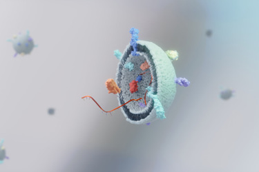
Extracellular vesicles (EVs) are non-replicating, lipid bilayer-enclosed particles naturally released by all cell types. These heterogeneous vesicles play a crucial role in intracellular communication and pathophysiological processes by delivering a wide range of biomolecules, including proteins, nucleic acids, lipids, and metabolites, making them promising candidates for therapeutic applications. Rapid analysis of EVs is essential, particularly for supporting clinical EV biomanufacturing platforms or studying their potential as carriers of nucleic acids (DNA or RNA) in relation to their parental cells. However, reliable size-based concentration measurements and specific marker-based characterization of EVs remain challenging with current analytical methods.
To address this, we developed a rapid analytical method using conventional flow cytometry, which demonstrated reliable accuracy, repeatability, linearity, and precision within a defined quantitation range. By utilizing a 405 nm Violet laser configuration, we were able to resolve nanoparticles ≥100 nm in size. Additionally, surface staining of EVs with established biomarkers enabled multiplexed characterization of EV phenotypes, showcasing the potential for advanced EV analysis.
Get unlimited access to:
Enter your credentials below to log in. Not yet a member of Outsourced Pharma? Subscribe today.
